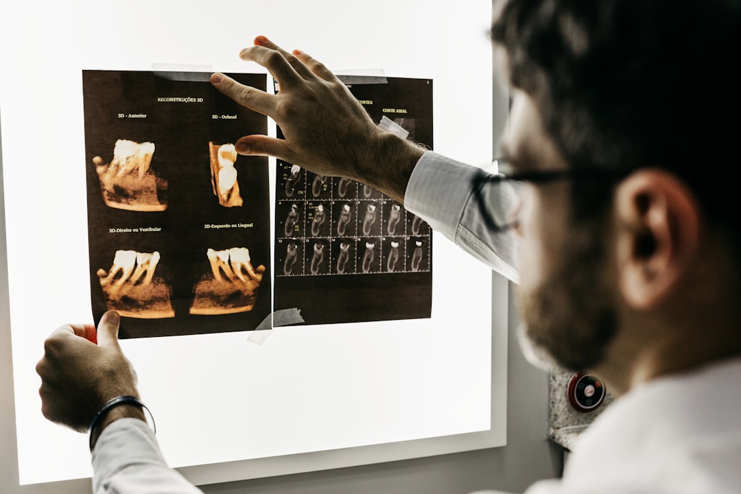
Cone Beam Computed Tomography (CBCT) scans are a type of medical imaging technology that uses a cone-shaped X-ray beam to create detailed 3D images of the teeth, jaw, and surrounding structures. Unlike traditional 2D dental X-rays, which provide a flat image of the oral cavity, CBCT scans offer a comprehensive view of the entire area, allowing for more accurate diagnosis and treatment planning. The CBCT scanner rotates around the patient’s head, capturing multiple X-ray images from different angles. These images are then reconstructed by a computer to create a 3D model of the oral and maxillofacial structures. This technology provides dentists and oral surgeons with a detailed view of the patient’s anatomy, allowing for more precise diagnosis and treatment planning.
CBCT scans are particularly useful for evaluating the bone structure, identifying dental abnormalities, and planning for dental implants and other oral surgeries. The high-resolution images produced by CBCT scans allow dental professionals to assess the quality and quantity of bone, detect pathology, and visualize the position of nerves and other vital structures. This level of detail is essential for accurate treatment planning and can help minimize the risk of complications during dental procedures. Additionally, CBCT scans can be used to evaluate the airway and temporomandibular joint (TMJ) disorders, providing valuable information for the diagnosis and management of these conditions. Overall, CBCT scans are a valuable tool for improving diagnostic accuracy and treatment outcomes in dentistry.
Key Takeaways
- CBCT scans provide 3D visualization of oral structures, allowing for comprehensive views of the patient’s anatomy.
- The enhanced precision of CBCT scans plays a crucial role in guided implant placement, improving the accuracy of the procedure.
- CBCT scans improve diagnostic capabilities, leading to more effective treatment planning and better patient outcomes.
- CBCT scans minimize radiation exposure, offering safety benefits for both patients and dental professionals.
- Utilizing CBCT scans can streamline treatment processes, leading to more efficient workflow and better patient care.
Improved Diagnostic Capabilities: How CBCT Scans Enhance Treatment Planning
CBCT scans offer significant advantages over traditional dental X-rays when it comes to diagnostic capabilities and treatment planning. The detailed 3D images produced by CBCT scans allow dental professionals to visualize the teeth, bone, nerves, and other structures from multiple angles, providing a more comprehensive understanding of the patient’s oral anatomy. This level of detail is particularly valuable for evaluating complex dental cases, such as impacted teeth, root fractures, and dental trauma. Additionally, CBCT scans can help identify pathology, such as cysts or tumors, that may not be visible on traditional X-rays. This enhanced diagnostic capability allows for earlier detection and intervention, potentially improving treatment outcomes for patients.
In addition to improving diagnostic accuracy, CBCT scans also play a crucial role in treatment planning for dental implants and other oral surgeries. By providing detailed information about the bone structure and surrounding anatomy, CBCT scans enable dental professionals to plan the precise placement of implants and design custom surgical guides. This level of precision is essential for ensuring the long-term success of dental implant procedures and minimizing the risk of complications. Overall, the improved diagnostic capabilities offered by CBCT scans have revolutionized the field of dentistry, allowing for more accurate diagnosis and treatment planning, ultimately leading to better outcomes for patients.
Enhanced Precision: The Role of CBCT Scans in Guided Implant Placement
One of the most significant advancements in dentistry facilitated by CBCT scans is the ability to perform guided implant placement with enhanced precision. Traditional implant placement relies on 2D imaging techniques, which may not provide a complete understanding of the patient’s bone structure and surrounding anatomy. In contrast, CBCT scans offer a detailed 3D view of the jawbone, allowing dental professionals to accurately assess bone density, volume, and quality. This information is crucial for determining the optimal location and angle for implant placement, as well as identifying any potential obstacles, such as nerves or sinus cavities.
Guided implant placement involves using the 3D images from CBCT scans to create a surgical guide that precisely outlines the position and angle for implant placement. This custom guide is fabricated based on the patient’s unique anatomy, ensuring accurate implant placement with minimal invasiveness. By utilizing CBCT scans for guided implant placement, dental professionals can achieve greater precision and predictability in their procedures, leading to improved long-term success rates for dental implants. Additionally, guided implant placement can reduce surgical time and minimize post-operative discomfort for patients. Overall, CBCT scans have revolutionized implant dentistry by providing a level of precision that was previously unattainable with traditional imaging techniques.
Minimizing Radiation Exposure: The Safety Benefits of CBCT Scans
| CBCT Scan Safety Benefits | Metrics |
|---|---|
| Reduced Radiation Exposure | 50-90% less radiation compared to conventional CT scans |
| Precise Imaging | High-quality 3D images with minimal distortion |
| Improved Diagnosis | Accurate visualization of dental and maxillofacial structures |
| Enhanced Safety | Lower risk of radiation-related health issues for patients |
While CBCT scans offer significant advantages in terms of diagnostic capabilities and treatment planning, concerns about radiation exposure have been raised regarding this imaging technology. However, it is important to note that CBCT scans are designed to minimize radiation exposure while still providing high-quality images for diagnostic purposes. Compared to medical CT scans, which use a fan-shaped X-ray beam and higher radiation doses, CBCT scans utilize a cone-shaped X-ray beam that delivers a lower radiation dose to the patient. Additionally, modern CBCT scanners are equipped with advanced technology that allows for dose optimization based on the specific imaging requirements.
Furthermore, dental professionals are trained to follow strict guidelines for radiation safety and dose optimization when performing CBCT scans. This includes using appropriate imaging protocols based on the patient’s age, size, and clinical indications, as well as employing shielding devices to minimize radiation exposure to non-targeted areas. By adhering to these guidelines and utilizing modern CBCT technology, dental professionals can ensure that patients receive the benefits of high-quality imaging while minimizing their radiation exposure. Overall, the safety benefits of CBCT scans lie in their ability to provide detailed diagnostic information with lower radiation doses compared to medical CT scans, making them a valuable tool in modern dentistry.
Comprehensive Views: How CBCT Scans Provide 3D Visualization of Oral Structures
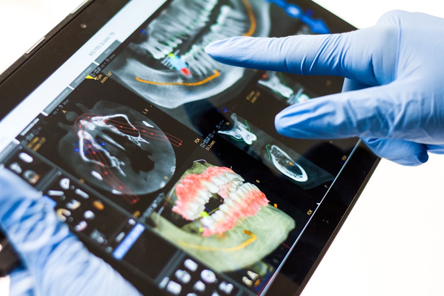
CBCT scans offer a level of comprehensive visualization that is unparalleled by traditional 2D dental X-rays. By capturing multiple X-ray images from different angles around the patient’s head, CBCT scanners create a 3D model of the oral and maxillofacial structures with exceptional detail. This allows dental professionals to visualize the teeth, bone, nerves, sinuses, and other anatomical structures from any perspective, providing a complete understanding of the patient’s oral anatomy. The ability to view these structures in 3D is particularly valuable for evaluating complex dental cases, such as impacted teeth, root fractures, or temporomandibular joint (TMJ) disorders.
In addition to providing comprehensive views of oral structures, CBCT scans also allow for precise measurements and virtual treatment planning. Dental professionals can use specialized software to analyze the 3D images and make accurate measurements of bone volume, density, and quality. This information is essential for planning dental implant procedures, orthognathic surgery, and other oral surgeries with a high level of precision. Furthermore, virtual treatment planning based on CBCT scans enables dental professionals to simulate procedures and anticipate potential challenges before performing them on patients. Overall, the comprehensive 3D visualization provided by CBCT scans has revolutionized the field of dentistry by offering a level of detail and accuracy that was previously unattainable with traditional imaging techniques.
Efficient Workflow: Streamlining Treatment Processes with CBCT Scans
In addition to improving diagnostic capabilities and treatment planning, CBCT scans also play a crucial role in streamlining treatment processes and enhancing workflow efficiency in dental practices. By providing detailed 3D images of the oral anatomy, CBCT scans enable dental professionals to make more accurate diagnoses and develop comprehensive treatment plans in less time. This can lead to more efficient patient care and reduced treatment times overall. Additionally, the ability to visualize complex dental cases in 3D allows for better communication between dental professionals and specialists, leading to more coordinated and streamlined care for patients.
Furthermore, CBCT scans can be integrated with digital impression systems and CAD/CAM technology to facilitate the design and fabrication of custom restorations, such as crowns, bridges, and dental implants. This integration allows for a seamless workflow from diagnosis to treatment delivery, reducing the need for multiple appointments and improving patient satisfaction. Additionally, by utilizing CBCT scans for guided implant placement and virtual treatment planning, dental professionals can optimize their surgical procedures and minimize post-operative complications. Overall, the efficient workflow facilitated by CBCT scans has transformed the way dental practices operate, leading to improved patient care and outcomes.
Patient Education and Engagement: Utilizing CBCT Scans to Enhance Communication and Understanding
CBCT scans have also proven to be valuable tools for patient education and engagement in dental practices. By visualizing the patient’s oral anatomy in 3D, dental professionals can effectively communicate complex diagnoses and treatment plans in a way that is easily understood by patients. This visual representation allows patients to see their oral structures from multiple angles and gain a better understanding of their condition and recommended treatment options. Additionally, by involving patients in the treatment planning process using CBCT images, dental professionals can empower them to make informed decisions about their oral health care.
Furthermore, CBCT scans can be used to demonstrate the potential outcomes of proposed treatments through virtual simulations. This allows patients to visualize the expected results before undergoing any procedures, leading to increased confidence and satisfaction with their treatment plans. Additionally, by actively involving patients in their care using CBCT images as educational tools, dental professionals can improve patient compliance and overall treatment outcomes. Overall, utilizing CBCT scans for patient education and engagement has proven to be an effective way to enhance communication between dental professionals and patients while promoting active participation in oral health care decisions.
In conclusion, Cone Beam Computed Tomography (CBCT) scans have revolutionized the field of dentistry by offering advanced diagnostic capabilities, enhanced precision in treatment planning, comprehensive visualization of oral structures in 3D, streamlined workflow efficiency in dental practices, minimized radiation exposure for patients’ safety benefits while providing valuable tools for patient education and engagement. The detailed 3D images produced by CBCT scans allow dental professionals to visualize the teeth, bone structure from multiple angles providing a more comprehensive understanding of the patient’s oral anatomy leading to improved diagnostic accuracy ultimately leading to better outcomes for patients.
In addition, the ability to accurately assess the position and orientation of teeth, evaluate the quality and quantity of bone for implant placement, and detect pathologies such as cysts, tumors, and fractures, has significantly improved the overall standard of care in dentistry. Furthermore, the integration of CBCT technology has allowed for more precise and minimally invasive surgical procedures, leading to reduced post-operative complications and faster recovery times for patients. Overall, CBCT scans have become an indispensable tool in modern dentistry, contributing to improved patient care, treatment outcomes, and overall practice efficiency.
FAQs
What is a CBCT scan in dental care?
A CBCT (cone beam computed tomography) scan is a specialized type of X-ray that provides detailed 3D images of the teeth, jaw, and surrounding structures. It is commonly used in dental care for treatment planning and diagnosis.
How is a CBCT scan different from traditional dental X-rays?
CBCT scans provide a more comprehensive view of the oral and maxillofacial structures compared to traditional dental X-rays. They offer detailed 3D images that allow for better visualization of the teeth, bone, nerves, and soft tissues.
What are the benefits of a CBCT scan in dental treatment?
CBCT scans can help dentists and oral surgeons accurately diagnose and plan treatment for various dental conditions, such as impacted teeth, dental implants, TMJ disorders, and orthodontic treatment. They also aid in identifying and assessing dental infections, tumors, and other abnormalities.
Is a CBCT scan safe for dental patients?
CBCT scans involve a lower radiation dose compared to medical CT scans, making them relatively safe for dental patients. However, pregnant women and children are typically advised to avoid unnecessary exposure to radiation, so the use of CBCT scans should be carefully considered in these cases.
How long does it take to get the results of a CBCT scan?
The results of a CBCT scan are typically available shortly after the scan is completed. The 3D images can be viewed and analyzed by the dentist or oral surgeon immediately, allowing for prompt treatment planning and decision-making.